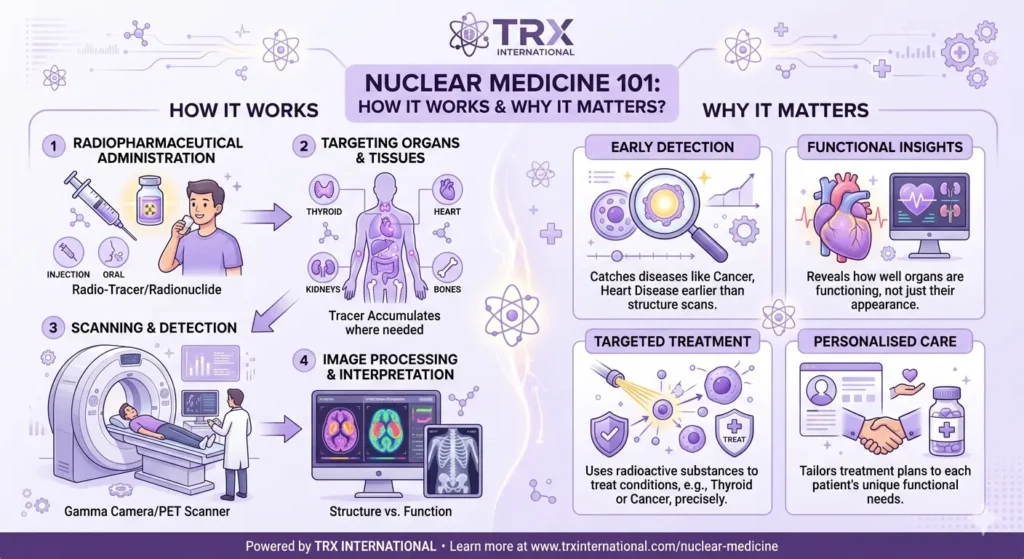
Nuclear Medicine 101: How It Works & Why It Matters?
Disclaimer: The information shared here is gathered from various sources and is intended for general awareness only, not as medical advice.
When you hear the words “nuclear” and “medicine” in the same sentence, your mind probably jumps to something out of a sci-fi film. The truth is far less dramatic and a whole lot more useful.
Nuclear medicine is one of the most fascinating branches of modern healthcare, blending physics, chemistry, and clinical care into a discipline that quite literally lets doctors see what’s happening inside your body at a molecular level.
So let’s break it down properly. Whether you’re curious about your upcoming scan, exploring a career in the field, or just trying to make sense of a doctor’s referral, this guide will walk you through everything you need to know about nuclear medicine.
What is Nuclear Medicine?
Nuclear medicine is a specialised area of medical imaging and treatment that uses tiny amounts of radioactive material to diagnose and treat a wide range of diseases. Unlike a standard x-ray or ct scan, which mostly show structure, nuclear medicine reveals function. It tells doctors how organs and tissues are actually performing in real time.
Think of it this way. An x-ray is like looking at a photograph of a car engine. Nuclear medicine imaging is like watching the engine run. Both have value, but they answer different questions about what’s going on under the hood.
The discipline sits at the intersection of radiology, oncology, cardiology, neurology, and endocrinology. It’s used everywhere from cancer detection to heart disease assessment, and it has quietly become one of the most important diagnostic tools available to modern healthcare.
Connect and Level Up Your Game
If you are interested in roles in nuclear space, reach out to the team at TRX International. We often have insights into upcoming outage needs and permanent staff positions before they hit the general job boards.
How Nuclear Medicine Differs from Other Imaging Studies?
| Feature | Nuclear Medicine | CT Scan | MRI | X-Ray |
|---|---|---|---|---|
| What it shows | Function and metabolism | Detailed structure | Soft tissue and structure | Bones and dense tissue |
| Technology used | Radioactive tracer + gamma camera or PET scanner | X-ray beams + computed tomography | Magnetic field + radio waves | X-ray beams |
| Radiation involved | Low to moderate | Moderate to high | None | Low |
| Best for | Cancer staging, thyroid, heart function, bone metastasis | Trauma, internal bleeding, tumors, lung issues | Brain, spine, joints, soft tissue | Fractures, chest, dental |
| Scan duration | 20 minutes to 1 hour | 5 to 15 minutes | 30 to 60 minutes | A few minutes |
| Contrast or tracer | Radiopharmaceutical injection | Iodine-based contrast (sometimes) | Gadolinium contrast (sometimes) | Usually none |
| Typical cost range | Moderate to high | Moderate | High | Low |
| Patient comfort | High, open scanner | High, quick procedure | Lower, enclosed and noisy | Very high |
Most people are familiar with traditional imaging like x-ray, ct, and mri. These technologies show what something looks like. Nuclear medicine, on the other hand, shows what something does. That distinction is huge.
A standard ct scan can spot a lump in your liver. A nuclear medicine scan can tell you whether that lump is metabolically active, which often determines whether it’s cancerous or benign. This is why oncology relies so heavily on nuclear imaging when staging tumors or tracking metastasis.
The information gathered from these imaging procedures helps a healthcare provider make decisions that genuinely change outcomes. Detecting disease earlier, choosing the right therapy, and monitoring response to treatment all become significantly more accurate with the help of molecular imaging.
Brief History of Nuclear Medicine
The roots of nuclear medicine stretch back to the 1930s when scientists first began experimenting with radioactive iodine to study thyroid function. By the 1950s, the field had grown enough that the society of nuclear medicine was formally established to support research, education, and clinical use.
Fast forward to today, and nuclear medicine procedures are performed millions of times every year around the world. From the development of the gamma camera to the rise of pet imaging and spect technology, the field has evolved at a pace few medical specialties can match.
What started as a niche research interest is now a cornerstone of diagnostic and therapeutic medicine, with new radiopharmaceutical compounds being approved regularly for both diagnosis and treatment.
Why Nuclear Medicine Matters in Modern Healthcare?
Here’s the thing. Nuclear medicine isn’t just about pretty pictures. It’s about answers. It helps clinicians diagnose and treat conditions that might otherwise go unnoticed until they’re far more advanced.
Cancer detection, heart disease evaluation, kidney function studies, brain disorders, and bone health assessments all benefit from nuclear medicine studies. The amount of radiation involved is generally low, and the diagnostic value is enormous, which is why most healthcare systems consider it essential.
For patients, this means earlier detection, better-targeted therapy, and often less invasive treatment paths. For clinicians, it means having a window into the body that no other imaging test can quite replicate.
How Nuclear Medicine Works? Tracers, Scanners, and Everything in Between
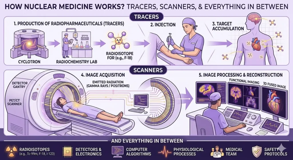
Now let’s get into the interesting part. How does nuclear medicine actually work? The short answer involves a radioactive tracer, a scanner, and some clever physics. The longer answer is worth understanding because it demystifies what can feel like a very intimidating process.
The Role of the Radioactive Tracer
At the heart of every nuclear medicine procedure is a substance called a radioactive tracer, sometimes referred to as a radiopharmaceutical. This is a small amount of radioactive material attached to a chemical compound that targets a specific organ, tissue, or biological process.
The tracer is usually delivered via intravenous injection, though some studies require oral administration or inhalation depending on what’s being examined. Once inside the body, the tracer travels to the area of interest. For example, radioactive iodine naturally accumulates in the thyroid, making it perfect for thyroid imaging.
As the tracer settles in, it gives off small amounts of energy in the form of gamma rays or positrons, which is what the scanner picks up to create the image. Without the tracer, there’s no signal, and without a signal, there’s no scan.
Common Imaging Modalities in Nuclear Medicine
There are several imaging modalities in nuclear medicine, but the two most common imaging modalities you’ll encounter are spect and pet. Let’s look at each one briefly.
Single photon emission computed tomography, or spect, uses a gamma camera that rotates around the patient to capture images from multiple angles. These are then reconstructed into 3D images. Spect is widely used for heart scan procedures, brain imaging, and bone studies.
Positron emission tomography, or pet scan technology, works a little differently. The tracer emits positrons that interact with electrons in the body, producing gamma rays detected by the pet scanner. Pet imaging is especially powerful in oncology because it shows metabolic activity, which often signals where cancer cells are most active.
Both spect and pet are sometimes combined with a ct scan to give doctors both functional and structural information in a single session. This hybrid approach has become standard in many nuclear medicine departments.
What Happens During a Nuclear Medicine Scan?
Most patients are surprised by how straightforward nuclear medicine scans actually are. There’s no claustrophobic tunnel like with an mri, and the procedure is generally painless aside from the brief injection.
After the tracer is administered, you’ll usually wait somewhere between 30 minutes and a few hours, depending on the type of scan. This waiting time allows the tracer to reach its target. Once it has, you lie still on a table while the scanner captures the images. The whole process typically takes anywhere from 20 minutes to an hour.
The technologist running the scan will guide you through every step. They monitor the equipment, position you correctly, and ensure the imaging procedures go smoothly from start to finish. Their role is hands-on and absolutely essential.
Build Your Nuclear Dream Team
Every unfilled role is a missed deadline. Top nuclear talent is scarce and getting scarcer. TRX International sources pre-vetted specialists globally so your projects stay on schedule and fully compliant.
Radiation Dose and Safety
| Source of Radiation | Typical Effective Dose (mSv) | Equivalent Context |
|---|---|---|
| Natural background radiation (per year) | 3.0 mSv | Baseline everyone receives |
| Chest X-ray | 0.1 mSv | About 10 days of background radiation |
| Dental X-ray | 0.005 mSv | A few hours of background radiation |
| Mammogram | 0.4 mSv | Around 7 weeks of background radiation |
| Bone scan (nuclear medicine) | 4.2 mSv | Roughly 1.4 years of background radiation |
| Cardiac SPECT scan | 9.0 mSv | Around 3 years of background radiation |
| PET/CT scan | 14.0 mSv | About 4.5 years of background radiation |
| Standard CT scan (abdomen) | 8.0 mSv | Around 2.7 years of background radiation |
| Radioactive iodine therapy | Higher therapeutic dose | Calibrated specifically per patient |
| Coast-to-coast flight (US) | 0.03 mSv | A few days of background radiation |
Naturally, anything involving the word radioactive raises eyebrows. So let’s address the elephant in the room. The radiation dose used in most nuclear medicine procedures is comparable to what you’d receive from a standard ct scan, and in many cases significantly less.
Radiation exposure to patients is carefully calculated to deliver just enough signal for accurate imaging while keeping the dose as low as reasonably achievable. Modern equipment, refined tracer chemistry, and improved imaging algorithms have all contributed to lower radiation exposure over the years.
For perspective, the amount of radiation from a typical nuclear medicine scan is roughly equivalent to what you’d absorb from natural background radiation over a year of normal living. It’s a small price for the diagnostic information you get back.
The Science Behind Image Formation
So how does all this turn into an actual image? When the tracer emits radiation inside your body, the gamma camera or pet scanner detects those emissions and translates them into digital signals. These signals are then processed by computers to create detailed images of the targeted organ or tissue.
The result is a map of biological activity. Bright spots usually mean high activity, while dark areas indicate low or no activity. Radiologists and nuclear medicine physicians then interpret these images, comparing them with normal patterns to identify abnormalities.
It’s a beautiful blend of physics and biology. The tracer behaves predictably because of its chemistry. The scanner captures emissions because of its physics. And the interpretation happens because of years of medical training and pattern recognition.
Where Nuclear Medicine Fits Alongside CT and MRI?
You might be wondering how nuclear medicine compares with CT and MRI. Each has its strengths. Ct excels at structural detail, mri offers excellent soft tissue contrast, and nuclear imaging brings functional information to the table.
In real-world practice, these technologies often complement each other. A patient with suspected cancer might get a ct first to spot a mass, an mri to characterise it further, and a pet scan to determine whether it’s metabolically active. Each piece of the puzzle adds clarity, and together they form a comprehensive picture.
This is why most major hospitals invest heavily in all three. No single modality has all the answers, but combined they cover almost every diagnostic question a clinician might have.
Diagnostic Applications: From Tumors to Thyroid Issues
Nuclear medicine plays a major role in diagnosing a wide range of conditions. In oncology, pet imaging is used to detect tumors, evaluate metastasis, and track response to treatment. The ability to spot metastatic disease early can change the entire course of care.
For thyroid conditions, radioactive iodine is used both diagnostically and therapeutically. It helps assess hyperthyroidism, identify thyroid cancer, and even treat overactive thyroid glands. The fact that the same tracer can both detect and treat disease is one of the most elegant features of nuclear medicine.
Other applications include heart scan procedures to evaluate coronary artery disease, bone scans to identify fractures or metastasis, kidney imaging to assess function, and brain scans to investigate dementia, epilepsy, and stroke.
Therapeutic Applications: Nuclear Medicine Therapy in Action
Nuclear medicine isn’t just diagnostic. It’s also therapeutic. Nuclear medicine therapy uses radionuclide compounds to deliver targeted radiation directly to diseased cells, sparing healthy tissue as much as possible.
Radioactive iodine therapy, for example, is the gold standard for treating certain types of thyroid cancer and hyperthyroidism. The thyroid naturally absorbs iodine, so when radioactive iodine is given, it concentrates in the thyroid and destroys problematic cells from the inside out.
Targeted radionuclide therapy has expanded dramatically in recent years, especially for prostate cancer, neuroendocrine tumors, and certain bone metastases that cause bone pain. These therapies often improve quality of life significantly when other options have run out.
Nuclear Medicine in Cardiology
Heart disease is another area where nuclear medicine shines. Cardiac spect studies use a tracer to evaluate blood flow to the heart muscle, helping cardiologists identify blockages, assess damage after a heart attack, and decide whether a patient needs further intervention.
Pet imaging of the heart provides even more detailed information about myocardial perfusion and viability. Sometimes red blood cells are labelled with a radioactive tracer to study cardiac function in motion, giving clinicians a dynamic look at how the heart pumps.
These studies are non-invasive, well-tolerated, and often replace the need for more aggressive diagnostic procedures. For patients with chest pain or known heart disease, nuclear cardiology can be a game-changer.
Nuclear Medicine in Neurology and Beyond
Brain imaging using nuclear medicine has opened up entirely new avenues for understanding conditions like Alzheimer’s, Parkinson’s, and epilepsy. Pet scans can detect changes in brain metabolism years before structural changes appear on a ct scan or mri.
Beyond the brain, nuclear medicine is used in pulmonary studies to detect blood clots in the lungs, in gastrointestinal studies to track bleeding or motility issues, and in renal studies to assess kidney function. The versatility is honestly astonishing once you start exploring the full scope.
There’s barely an organ system in the body that doesn’t benefit from some form of nuclear medicine imaging or therapy at this point. That’s why the field continues to grow year after year.
Looking to hire nuclear professionals or explore nuclear career opportunities?
TRX International connects world-class talent with critical roles across the global nuclear industry. Visit trx-international.com or get in touch with the team to start the conversation.
Preparing for a Nuclear Medicine Procedure
If you’re scheduled for a nuclear medicine scan, preparation is usually minimal but important. Your healthcare provider will give you specific instructions based on the type of study being performed.
For some scans, you may need to fast for a few hours beforehand. For others, you might be asked to stop certain medications temporarily. Hydration is often encouraged because it helps the body process and excrete the tracer more efficiently after the imaging test is complete.
It’s also worth letting your provider know if you’re pregnant, breastfeeding, or have any allergies. These factors don’t always rule out nuclear medicine, but they do influence how the procedure is approached.
After the Scan: What to Expect?
Once the imaging is complete, you can usually go about your day as normal. The radioactive tracer wears off relatively quickly, and the small amount of radiation it produces dissipates within hours to a few days depending on the specific compound used.
You’ll be advised to drink plenty of water to help flush the tracer out of your system. Some patients are told to avoid close contact with pregnant women or young children for a short period, especially after certain therapeutic procedures, but this is precautionary rather than alarming.
Results are typically available within a day or two, depending on the complexity of the scan and how busy the nuclear medicine department is. Your healthcare provider will then discuss what the imaging studies revealed and what the next steps might be.
The People Behind Nuclear Medicine
Nuclear medicine isn’t a one-person operation. It’s a team effort involving nuclear medicine physicians, technologists, medical physicists, radiopharmacists, nurses, and support staff. Each plays a critical role in making sure the diagnostic and therapeutic procedures run safely and accurately.
Nuclear medicine physicians are specialists who interpret the scans and oversee patient care. Technologists handle the day-to-day operation of imaging equipment and patient interaction. Medical physicists ensure radiation safety and quality control, while radiopharmacists prepare the radioactive compounds with extraordinary precision.
This multidisciplinary structure is one of the things that makes nuclear medicine such an interesting field to work in. There’s a place for almost every kind of skill set, from clinical care to engineering to chemistry.
Career Pathways in Nuclear Medicine
If you’re reading this because you’re considering a career in the field, the good news is that nuclear medicine offers a remarkable variety of pathways. You can train as a nuclear medicine technologist, pursue medical school and specialise as a nuclear medicine physician, or focus on the technical and physics side as a medical physicist.
Other pathways include radiopharmacy, which involves preparing the radioactive compounds, and nursing roles specialised in nuclear medicine and oncology care. There’s also growing demand for engineers and software developers who work on imaging equipment and image processing algorithms.
The demand for skilled professionals continues to grow as nuclear medicine expands. And while training can be rigorous, the work is genuinely rewarding because you’re directly contributing to patient outcomes in a tangible way.
Why the Nuclear Industry is Growing?
Behind every nuclear medicine procedure is a complex supply chain that depends on the broader nuclear industry. The radionuclide isotopes used to make tracers are produced in nuclear reactors and cyclotrons, often in just a handful of facilities worldwide.
This means the future of nuclear medicine is closely tied to investments in nuclear infrastructure, including new reactor designs, isotope production facilities, and supply chain resilience. Governments and private companies are increasingly recognising this connection and ramping up investment accordingly.
The intersection of nuclear energy, healthcare, and advanced manufacturing has created a booming sector with diverse career opportunities. From reactor operators to logistics specialists, from regulatory affairs to research scientists, the people who make nuclear medicine possible work across an incredibly wide range of disciplines.
Recruitment and Talent in Nuclear Medicine
This is where specialised recruitment becomes critical. The nuclear industry, including nuclear medicine, has a serious skills gap. The workforce is ageing, demand is rising, and the technical expertise required is significant. Hiring the right people is no longer just an HR function. It’s a strategic priority.
That’s where firms like TRX International come in. As a leading nuclear recruitment agency, TRX International specialises in connecting top-tier talent with employers across the nuclear sector, including roles that support nuclear medicine, isotope production, regulatory affairs, and clinical operations.
Whether a hospital is looking for a senior nuclear medicine physician, a reactor facility needs a radiopharmaceutical specialist, or a startup is building the next generation of pet imaging technology, having a recruitment partner who understands the nuclear industry inside out makes all the difference.
The Future of Nuclear Medicine and Molecular Imaging
The field is evolving rapidly. Advances in molecular imaging, artificial intelligence, and personalised medicine are reshaping what’s possible. New radiopharmaceutical compounds are being developed every year, targeting more specific biological pathways with greater precision than ever before.
Theranostics, a hybrid approach that combines diagnostics and therapy using the same molecular target, is one of the most exciting developments. It allows clinicians to identify exactly where disease is and then deliver treatment precisely to that location, all using nuclear medicine principles.
As technology continues to improve and access expands, nuclear medicine will likely play an even bigger role in everything from early cancer detection to personalised cardiac care. The next decade promises to be transformative.
Common Misconceptions About Nuclear Medicine
There are plenty of myths floating around about nuclear medicine, and most of them aren’t true. Let’s clear a few up.
First, nuclear medicine doesn’t make you radioactive in any meaningful way. The tracers used clear from your body quickly, and the amount of radiation involved is small. Second, you won’t glow in the dark or set off airport security. Third, the procedures aren’t painful, and most patients tolerate them very well.
It’s also worth pointing out that nuclear medicine is different from radiation therapy used in cancer treatment, although there’s some overlap with targeted radionuclide therapy. They share underlying principles but serve different clinical purposes most of the time.
When You Might Need Nuclear Medicine?
Your healthcare provider might recommend a nuclear medicine scan for all sorts of reasons. Common scenarios include investigating unexplained bone pain, evaluating thyroid function, assessing heart disease, staging cancer, or monitoring response to treatment.
Sometimes the recommendation comes after other imaging like a ct scan or mri has raised questions that only functional imaging can answer. Other times, nuclear medicine is the first-line investigation, especially for conditions like hyperthyroidism or certain cancers where metabolic activity is the key piece of information.
If you’re ever unsure why a particular scan has been ordered, ask your healthcare provider. Understanding the reason behind a test usually makes the whole experience less stressful and helps you engage more meaningfully with your care.
Insurance, Access, and Cost Considerations
Nuclear medicine procedures are widely covered by insurance and national health systems, especially when ordered for established clinical reasons. That said, costs can vary depending on the country, the specific scan, and the facility.
Pet scans tend to be among the more expensive nuclear medicine studies due to the complexity of the equipment and the cost of the tracer. Spect scans and standard nuclear medicine procedures are usually more affordable. In many regions, access has improved significantly over the past decade as more facilities have invested in this technology.
If cost is a concern, talk to your healthcare provider or insurance company. There are often options to make these critical diagnostic and therapeutic procedures accessible.
Quick-Reference Cheat Sheet: Nuclear Medicine Essentials
| Concept | What It Means |
|---|---|
| Tracer | Small amount of radioactive material used to create images |
| Spect | Single photon emission computed tomography, used for heart, brain, and bone |
| Pet | Positron emission tomography, used heavily in oncology |
| Radiopharmaceutical | The full compound combining tracer and targeting agent |
| Gamma Camera | Device that detects gamma rays from the tracer |
| Radionuclide | Radioactive isotope used as the energy source in tracers |
| Theranostics | Combined diagnostic and therapeutic nuclear medicine approach |
Frequently Asked Questions
Is Nuclear Medicine Safe?
Yes. The amount of radiation involved is carefully controlled and generally comparable to or less than a standard ct scan. The diagnostic and therapeutic value far outweighs the small risk associated with radiation exposure.
How long does a Nuclear Medicine scan take?
Most scans take between 20 minutes and an hour for the imaging itself, though waiting time after the tracer injection can extend the total visit to a few hours.
Can pregnant women have nuclear medicine scans?
Generally, nuclear medicine procedures are avoided during pregnancy unless absolutely necessary. Always inform your healthcare provider if you are or might be pregnant.
What’s the difference between a pet scan and a ct scan?
A ct scan shows structural anatomy using x-ray technology. A pet scan shows metabolic and functional activity using a radioactive tracer. They’re often combined in modern hybrid scanners for the most complete picture.
How is nuclear medicine therapy different from chemotherapy?
Chemotherapy uses drugs that affect cells throughout the body. Nuclear medicine therapy uses radionuclide compounds that target specific cells based on biological behaviour, often resulting in fewer systemic side effects.
Do I need to avoid people after a nuclear medicine scan?
For most diagnostic scans, no special precautions are needed. After certain therapeutic procedures, especially radioactive iodine therapy, you may be asked to limit close contact with others for a short period.
Who interprets the results?
Nuclear medicine physicians are the specialists trained to interpret these scans. They work closely with your referring doctor to ensure the findings are integrated into your overall care plan.
Final Thoughts
Nuclear medicine sits quietly at the heart of modern healthcare, doing extraordinary work without the fanfare it probably deserves. From detecting cancer early to treating thyroid disease to evaluating heart function, this field touches more lives than most people realise.
For patients, understanding what nuclear medicine is and how it works can take a lot of the anxiety out of an upcoming scan. For professionals, it’s a field full of opportunity, innovation, and meaningful impact. And for the broader nuclear industry, it’s a reminder that nuclear technology has applications far beyond energy generation.
If you’re exploring opportunities in this space, whether as a clinician, technologist, scientist, or operations professional, the demand for skilled people has never been higher.
Specialist recruiters like TRX International are well-positioned to help match talent with the right opportunities, ensuring the future of nuclear medicine remains bright, well-staffed, and ready for whatever comes next.
Nuclear medicine is, in the end, a beautiful example of human ingenuity. It takes something as raw as radioactivity and turns it into healing, insight, and hope. That’s worth understanding, supporting, and investing in.